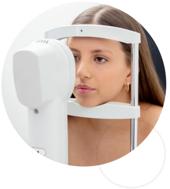
Equipped with the latest in digital imaging devices.
At Vision Camberwell, we recommend a comprehensive eye examination every two years to ensure optimal vision and eye health. These thorough assessments, lasting 30 minutes to an hour, may include pupil dilation to evaluate the macula, retina, and optic nerve. Our exams address your vision needs, providing tailored advice on spectacles or contact lenses, testing eye coordination and focusing, monitoring for conditions like cataracts, glaucoma, and macular degeneration, and measuring intra-ocular pressure. Advanced imaging, such as digital photography or 3D scans of the retina and optic nerve, further supports precise diagnoses and personalized care.
Advanced Technology and Equipment in Optometry
At our clinic, we are committed to delivering exceptional eye care by utilizing cutting-edge technology and equipment. These advanced tools allow us to provide comprehensive assessments, accurate diagnoses, and tailored treatment plans for a variety of ocular conditions. Below is an overview of the key technologies we use.
Optical Coherence Tomography (OCT)
Optical Coherence Tomography (OCT) is a non-invasive imaging method that generates high-resolution cross-sectional visuals of the retina and optic nerve. This advanced technology is critical for the early detection and management of eye diseases, including:
- Macular Degeneration: Visualizes changes in retinal layers.
- Glaucoma: Monitors optic nerve health.
- Diabetic Retinopathy: Detects retinal swelling or bleeding.
The integration of OCT-Angiography (OCT-A) further enables the examination of retinal blood vessels without the need for dye injections, providing unparalleled insights into ocular health.
Corneal Topography and Tomography
Corneal topography creates a detailed map of the cornea’s surface curvature, while corneal tomography goes deeper, assessing the cornea’s front and back surfaces as well as its thickness. These technologies are essential for:
- Contact Lens Fittings: Ensures precise fitting for rigid and specialty lenses.
- Keratoconus Management: Identifies and tracks corneal abnormalities.
- Refractive Surgery Planning: Evaluates suitability for procedures like LASIK.
Retinal Imaging
Digital retinal imaging captures high-resolution images of the retina, macula, and optic nerve. This allows for the detection of subtle changes in eye health over time. It is vital for monitoring conditions such as:
- Glaucoma: Tracks optic nerve damage.
- Macular Degeneration: Identifies drusen or pigment changes.
- Diabetic Retinopathy: Detects early signs of vascular damage.
Slit-Lamp Biomicroscopy
The slit lamp offers a highly magnified, illuminated view of the eye’s anterior structures, such as the cornea, iris, and lens. This technology is pivotal for:
- Diagnosing conditions like cataracts, corneal ulcers, and dry eye.
- Fitting specialty contact lenses.
- Monitoring ocular surface health.
Axial Length Measurement
Axial length measurement is crucial in managing myopia, as elongation of the eye increases the risk of complications such as retinal detachment and glaucoma. By monitoring axial length, we can effectively evaluate the progress of myopia control treatments, including orthokeratology and atropine therapy.
Visual Field Testing
Visual field testing assesses peripheral vision, playing a key role in detecting and monitoring:
- Glaucoma: Identifies early vision loss.
- Neurological Disorders: Evaluates visual field changes due to brain conditions.
- Driver Licensing: Ensures compliance with visual standards.
Automated perimetry provide precise measurements of vision sensitivity across the visual field.
Anterior Segment Imaging
Anterior segment imaging provides a 3D view of the eye’s front structures, aiding in the diagnosis and treatment of conditions such as:
- Dry Eye Syndrome: Evaluates tear film and ocular surface.
- Contact Lens Fitting: Assesses lens positioning and fit.
- Surgical Planning: Prepares for cataract and refractive surgeries.
Pachymetry
Pachymetry measures corneal thickness, essential for diagnosing and managing conditions such as:
- Keratoconus: Monitors progression.
- Glaucoma: Assesses risk factors based on corneal thickness.
Why Choose Us?
Our investment in state-of-the-art diagnostic equipment ensures that you receive the highest standard of care. By combining advanced technology with personalized treatment, we aim to protect and enhance your vision for years to come.
Contact us today to learn more or schedule your comprehensive eye examination.


